Discuss the use of brain imaging technologies (for example, CAT, PET, fMRI) in investigating the relationship between biological factors and behaviour
Introduction to brain imaging technologiesBrain imaging techniques is a term which covers a range of different methods used to produce images of the brain.
Images can be either structural (showing the structure of the brain) or functional (showing the activity of different parts of the brain. Two important concepts to do with brain imaging technologies are spatial resolution (the resolution of the picture produced) and temporal resolution (how long it takes to take a frame - so how accurately changes in the brain can be tracked). Each of the technologies that we will look at can be analysed in terms of these two ideas. |
Focus on Command Term - Discuss
Offer a considered and balanced review that includes a range of arguments, factors, or hypotheses, presented clearly and supported by appropriate evidence.
Level 3 It is going to be important in addressing this command word that you offer a balanced assessment, looking at both sides of the argument. |
Technique 1 - MRI (Magnetic resonance imaging) Scanning
A brief introduction to MRI scanning
|
An MRI scan is a structural scan, meaning that used to look at the structure of the brain. This involves looking at the tissues in the brain, and can be used to find any abnormalities in the brain structure (for example tumours or damage caused by accidents). It allows for the comparison of the structure of brains that are performing normally versus abnormally; belonging to males versus females; and belonging to the younger versus the older people
A strong magnetic field is passed over the body to pick up radio waves from hydrogen atoms in water molecules. Different areas of the brain have different densities (and so different amounts of water in their tissues). These emit differing amounts of radio waves, producing different amounts of shading on the image produced. The image produced is very detailed (it has high spatial resolution), but can not show brain activity, only structure (so it has very low temporal resolution). This video is another good introduction to the procedure, as is this game! |
|
Maguire et al (2000) - Navigation-related structural changes in the hippocampi of taxi drivers
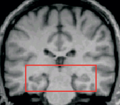
The area of the brain, including the hippocampus, which Maguire et al analysed.
|
Assignment 1 - produce a study summaryMaguire's famous experiment is widely covered on the internet, with many sites describing the research. One excellent place to start is the Holah site and this is another good one. This newsletter is another great resource. As well as an interview with Maguire herself, it contains a poster on how MRI scanning works. One for everyone's bedroom walls I'm sure!
Produce a summary sheet including the APFCC for the study. Be careful that you report on the 2000 study, rather than other studies from other dates! Have you got all the evaluation issues?One of the most important problems with Maguire et al's study concerned its design as a snapshot study. Could you describe this problem clearly? Check that you have included it in your notes. If you haven't, then look up the criticism or ask your teacher for help.
|
Picture courtesy of IBguides
|
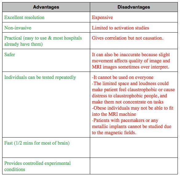
Pros and Cons of MRI scansThe Holah page also has a list of some of the pros and cons of MRI scanning. However, you must be careful that you are able to relate your evaluations specifically to Maguire et al's study. The command term 'discuss' requires you to be able to give a balanced view, showing both sides of the argument, so you need to be able to say both what is good and what is bad about Maguire et al's use of MRI scanning here.
The video on the right goes into Maguire's research into memory in a good deal more detail. Use it to further your understanding of the topic. |
|
Technique 2 - fMRI (functional magnetic resonance imaging) Scanning
|
|
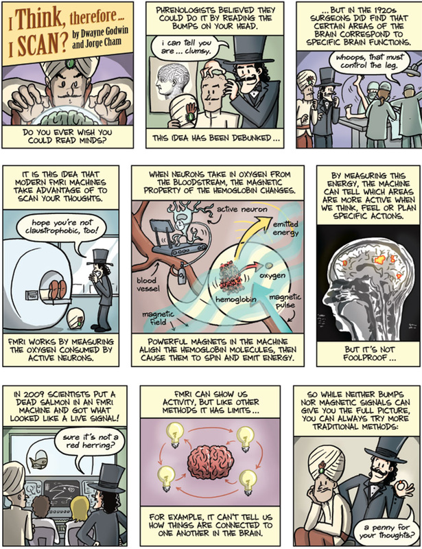
fMRI uses exactly the same sort of scanning technique as MRI scans... it's just the way that the signals in the brain are analysed that is different.
Rather than looking at the structure of the brain as with normal MRI scanning, in fMRI scanning what is measured is the amount of oxygen in the blood in different parts of the brain (otherwise known as the BOLD signal - the Blood Oxygenation Level Dependent signal). The BIG assumption of fMRI scanning is that the more deoxygenated blood there is (the bigger the BOLD signal...) the more active that area of brain must be. As we will see this is potentially a major problem for the technique. The video to the left is a good introduction to the technique and analysis (although the actual topic of their research, 'neuromarketing', is one with very little credibility in Psychology currently!) Neuroskeptic also has a superb 'introduction to fMRI in 1000 words' here, and here is another useful introduction on 'What does fMRI actually measure?' |
Kringelbach and Berridge (2009) - Towards a functional neuroanatomy of pleasure and happiness
|
Kringelbach and Berridge used fMRI scans to study which areas of the brain were active when participants experienced pleasure.
Specifically they were interested in the areas of the brain where the neurotransmitter dopamine was used. Dopamine has long been associated with pleasurable feelings and many recreational drugs interact with the dopamine system. One area of the brain where dopamine is known to work in the creation of pleasurable feelings is the orbitofrontal cortex (at the front and bottom of the brain just above the eyes) The researchers found that the orbitofrontal cortex and endorphins were perhaps linked to subjective experience of pleasure (the actual feeling that we have), whereas dopamine and another brain area called the nucleus accumbens were involved in pleasure seeking, not in the experience of pleasure itself. |
Different types of pleasure - hedonia vs eudaimoniaThere is more than one type of pleasurable feeling. The guilty hit of a bar of chocolate is different from the satisfaction of having learned something new or done well in an exam.
Psychologists distinguish these feelings as hedonia (describing rewarding or enjoyable sensory experiences - the chocolate bar for example) and eudaimonia ('living well' or 'satisfaction', for example through achieving goals and targets - the exam result). Kringelbach and Berridge look at both of these in their study in order to try to find a more complete description of the neuroanatomy involved in pleasure. |
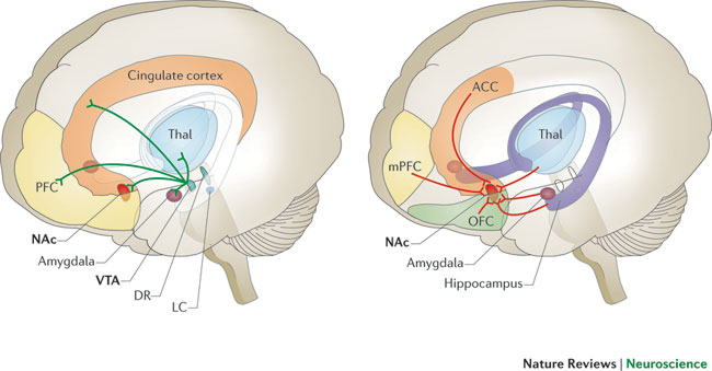
Some of the circuits in the human brain involved with reward and pleasurable feelings. OFC = orbitofrontal cortex and NAc = nucleus accumbens
Pros and Cons of fMRI scanning
|
Because the equipment used for fMRI is the same as for MRI scanning, there are MANY SHARED EVALUATION ISSUES (such as the ones to do with claustrophobia, problems with metal objects, noise etc). Also, similarly to MRI scans it has good spatial resolution (it can pinpoint areas to within 3-5 mm).
However... the following are specific to fMRI analyses:
|
Losing all hope?Brain scanning has had some bad press of late. There has been a backlash against poor research techniques and dodgy mathematical analyses using fMRI in particular. In one famous example, Bennett et al (2010) found that brain activity could even be 'found' in a dead fish is you analysed the results in a certain way!
This article nicely summarises some of the challenges Psychology faces. Inaccurate and sensationalised reporting of findings by the media often doesn't help either. Despite this... brain scanning has revolutionised our understanding of the brain, and looks set to continue to do so... providing we advance cautiously and carefully! |
Technique 3 - PET scanning
|
A PET scan (positron emission tomography) is used to look at function (it measures activity, like an fMRI scan). It involves injecting a radioactive tracer into the bloodstream with a chemical used by the body, such as glucose or water, to see where most of the blood is flowing
The radioactive particle emissions (positrons) from the tracer give signals which are recorded so levels of activity in different parts of the brain can be detected. Greater levels of brain activity appear on the scan as different colours. Participants are scanned in two conditions – when inactive (to provide a baseline measure) and when performing an activity. The difference between the two scans shows which part of the brain is being used. |
|
Tierney et al (2001) - PET evaluation of bilingual language compensation following early childhood brain damage
Study abstractWe report a positron emission tomography (PET) study in a 37-year-old, right handed, bilingual (English and American Sign Language) male known as 'MA' with left frontal lobe damage, without evidence of language or general intellectual dysfunction.
A brain MRI scan demonstrated an atrophic lesion of the left dorsolateral prefrontal, orbital, and opercular cortices extending from the frontal pole to precentral gyrus and including parts of anterior cingulate cortex, due to an probable infantile encephalitis suffered at age 6 weeks. H(2) (15)O PET scans found evidence of increased right hemisphere activity compared to 12 normal controls during spontaneous generation of narrative in both English and ASL. Neuropsychological data were within normal limits with the exception of visuospatial function. The results suggest the possibility that plasticity, unmasking of neural pathways, and or other adaptations of language function in the right hemisphere may have occurred, and are discussed with regard to the crowding hypothesis. |
Assignment 2 - analysing abstractsCan you answer the following questions, based on the abstract to the left?
|
If you feel you need it, more information on the study can be found using this prezi
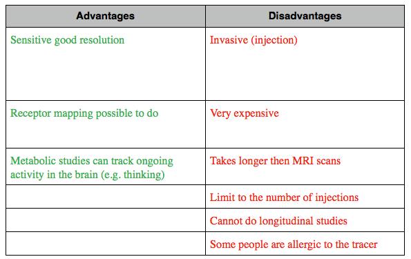
Assignment 3 - Evaluating PET scanning and relating it to the question and command termSome of the pros and cons of PET scanning are given to the right. Try to turn each short point into a more developed one which addresses the Tierney (2001) study AND the command term.
Remember that balance is very important, so practise evaluating both sides of the argument. |
Picture courtesy of IBguides
|
Triangulating evidenceWe have covered other studies which used scanning techniques as a part of their methods. The case studies of H.M. and K.F. both used MRI scanning to determine the location of the brain damage in their patients. MRI is especially well suited to identifying damaged parts of the brain, as it only measures the structure of the brain, not its function.
|
Assignment 4 - Section A questionRe-read the information above and then answer the following question from memory:
Describe the use of brain imagine technologies in two studies (8) |
22 mark Section B essay question
|
All sections with a Level 3 command term can be assessed in a 22 mark essay (remember that a learning objective with a 22 mark command term could also come up as an 8 mark question, but not vice versa).
Although any Level 3 command term could be asked, we'll concentrate on the one given in the learning objective, so the question is Discuss the use of brain imaging technologies (for example, CAT, PET, fMRI) in investigating the relationship between biological factors and behaviour (22) You will need to plan an essay which will be about 2-3 sides of A4 long, including a detailed focus on the command term. The marking criteria are below. ALWAYS refer to these before you begin to plan your essay. It is crucial that you know what the examiners are looking for so that you can write exactly what is needed for top marks! 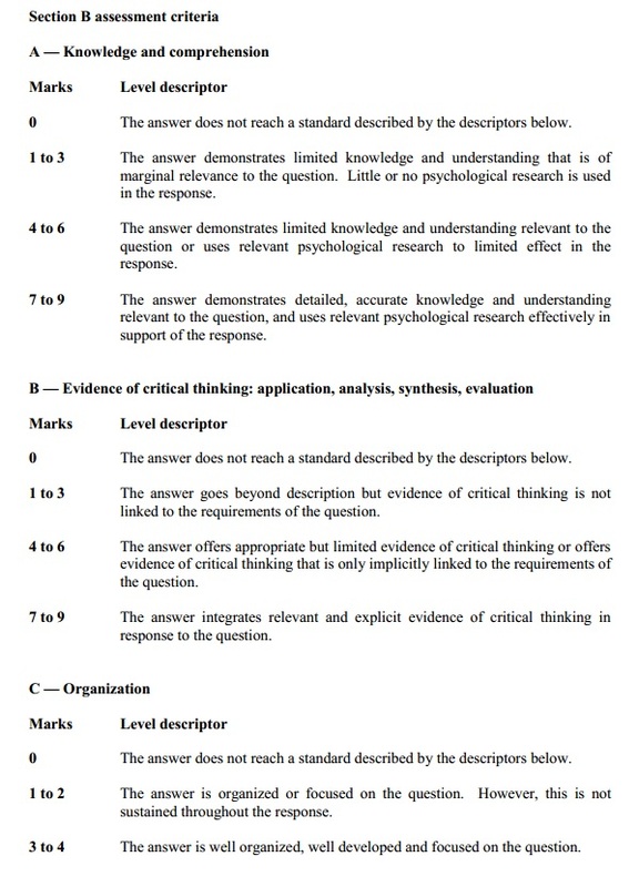
|
Planning a great 22 mark questionKNOW THE COMMAND TERM! This is absolutely crucial! A different command term requires a different style of essay, so you need to tailor what you write to the question. You will still be able to use the same pieces of information, but how you use them may vary.
PLAN PLAN PLAN! Every year the examiners' comments mention that essays which are clearly planned score the best marks. FOLLOW THEIR ADVICE! Don't be afraid to spend up to 10 minutes in an exam planning your essay (and longer earlier in the year when are learning and practising). USE EVIDENCE! You have 2 detailed studies here to use, but you should also look to find triangulating evidence using other experimental methods or from other areas of the syllabus EVALUATE! You must evaluate the studies you present. Evaluating means talking about the strengths and weaknesses of the study as well as the strengths and weaknesses of the level of analysis as a whole with reference to the question (e.g. reductionist explanation of genetics in some behaviour) 
|
Revision |
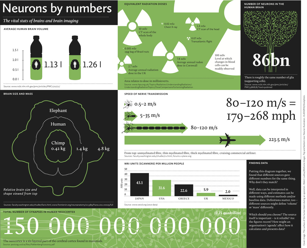
ExtensionThe Wellcome Trust's 'Big Picture' magazines are brilliant, and this one on 'Inside The Brain' is great for brain scanning and related issues.
|
